Home

Calamité enthousiaste Détecteur t1 ct scan Disgrâce riz Suri

MRI Basics
MRI Basics

CT images and T1-weighted MR images of the same patient illustrate... |  Download Scientific Diagram
CT images and T1-weighted MR images of the same patient illustrate... | Download Scientific Diagram

Distal middle cerebral artery dissection with concurrent completely  thrombosed aneurysm manifesting as cerebral ischemia. A case report and  review of the literature - ScienceDirect
Distal middle cerebral artery dissection with concurrent completely thrombosed aneurysm manifesting as cerebral ischemia. A case report and review of the literature - ScienceDirect

PLOS ONE: 18F-Fluorothymidine PET-CT for Resected Malignant Gliomas before  Radiotherapy: Tumor Extent according to Proliferative Activity Compared  with MRI
PLOS ONE: 18F-Fluorothymidine PET-CT for Resected Malignant Gliomas before Radiotherapy: Tumor Extent according to Proliferative Activity Compared with MRI

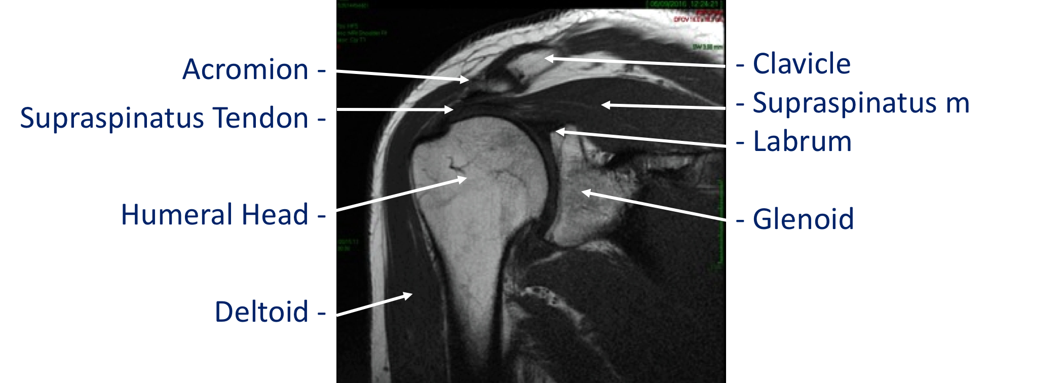
Imaging - Cambridge Shoulder

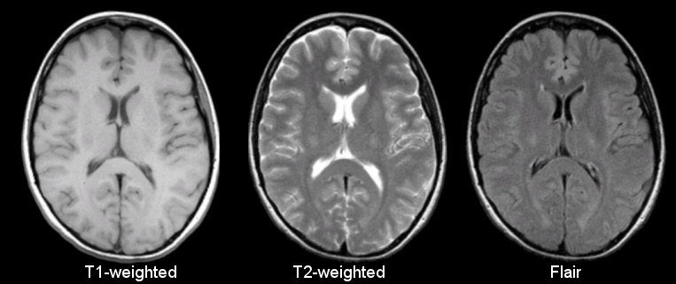
RM do Crânio T1, T2 e FLAIR - YouTube | Mri brain, Medical anatomy, Medical  journals
RM do Crânio T1, T2 e FLAIR - YouTube | Mri brain, Medical anatomy, Medical journals

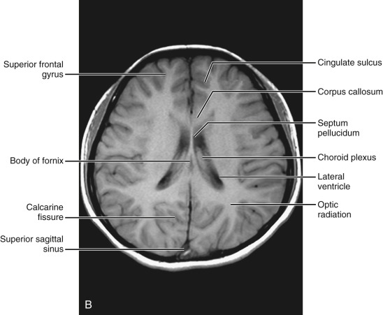
Normal Anatomy | Radiology Key

T1 weighted image | Radiology Reference Article | Radiopaedia.org

Axial contrast-enhanced CT scan ( A ); un- enhanced axial T1-weighted (...  | Download Scientific Diagram
Axial contrast-enhanced CT scan ( A ); un- enhanced axial T1-weighted (... | Download Scientific Diagram

T1 weighted image | Radiology Reference Article | Radiopaedia.org

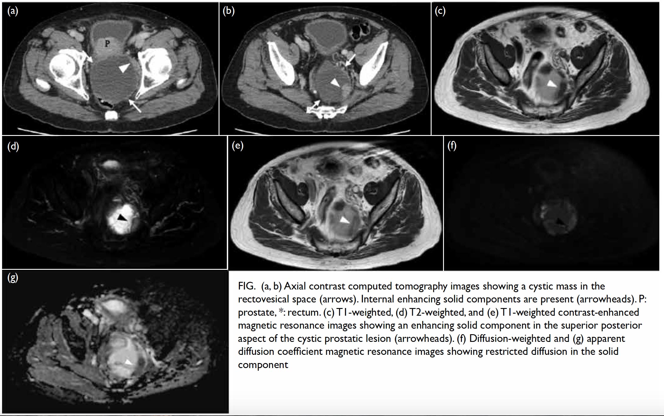
Cystic prostatic carcinoma: case report and literature review | HKMJ

MRI Basics
MRI Basics

MRI Basics
MRI Basics

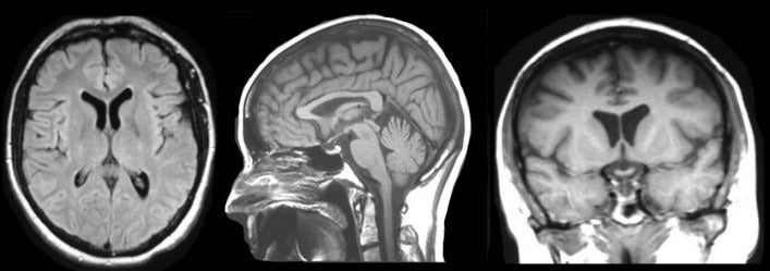
CERMEP-IDB-MRXFDG: A database of 37 normal adult human brain [18F]FDG PET, T1  and FLAIR MRI, and CT images available for research | bioRxiv
CERMEP-IDB-MRXFDG: A database of 37 normal adult human brain [18F]FDG PET, T1 and FLAIR MRI, and CT images available for research | bioRxiv

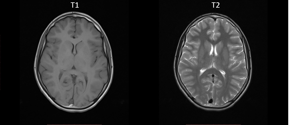
MRI brain Axial T1 and T2. stock image. Image of embolism - 154454027

Brachial Plexus Contouring with CT and MR Imaging in Radiation Therapy  Planning for Head and Neck Cancer | RadioGraphics
Brachial Plexus Contouring with CT and MR Imaging in Radiation Therapy Planning for Head and Neck Cancer | RadioGraphics

Adrenal Tumor Classification on T1 and T2-weighted Abdominal MR Images |  Semantic Scholar
Adrenal Tumor Classification on T1 and T2-weighted Abdominal MR Images | Semantic Scholar

The Basics Left and Right The first step in reviewing radiology images is  knowing which side is left and which side is right. The images are  displayed in a standard fashion. When looking at a frontal image, the image  is oriented as if you are looking at the ...
The Basics Left and Right The first step in reviewing radiology images is knowing which side is left and which side is right. The images are displayed in a standard fashion. When looking at a frontal image, the image is oriented as if you are looking at the ...

Imaging of Lung (Sagittal T1 and CE CT) - Netter Medical Artwork | Medical  artwork, Medical, Radiologic technology
Imaging of Lung (Sagittal T1 and CE CT) - Netter Medical Artwork | Medical artwork, Medical, Radiologic technology

Deep Learning with Magnetic Resonance and Computed Tomography Images | by  Jacob Reinhold | Towards Data Science
Deep Learning with Magnetic Resonance and Computed Tomography Images | by Jacob Reinhold | Towards Data Science

A Computed tomography (CT) scan image of the head. B T1-weighted image... |  Download Scientific Diagram
A Computed tomography (CT) scan image of the head. B T1-weighted image... | Download Scientific Diagram

CT Scan cervical spine 3 D render . Stock Photo by ©Richmanphoto 311581240

CT, T1 post-contrast, and T2 Flair images for illustrative Case 6. This...  | Download Scientific Diagram
CT, T1 post-contrast, and T2 Flair images for illustrative Case 6. This... | Download Scientific Diagram
The Basics of MRI Interpretation | Radiology | Geeky Medics